News
Digital Subtraction Angiography: A Comprehensive Overview
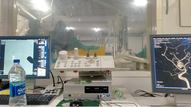
Digital Subtraction Angiography subtracts pre-contrast from post-contrast images for vascular imaging clarity. It displays stenosis, aneurysms, and arteriovenous malformations in blood arteries or vessels with low background interference. Consequently, it provides accurate, real-time guidance during interventional treatments to decrease risks and increase results. Read this article to learn more about Digital Subtraction Angiography!

Understanding Digital Subtraction Angiography
How to Define DSA?
As noted earlier, Digital Subtraction Angiography increases blood vessel visibility while removing pre- and post-contrast images. Therefore, vascular structures are isolated from surrounding tissues in the final image. DSA can find stenosis, aneurysms, and arteriovenous malformations.
How Does DSA Work?
DSA acquires a series of baseline and mask images without any contrast agent. Then, a contrast medium is injected into the bloodstream, and subsequent images are taken. The software subtracts the mask images from the contrast-filled images and eliminates non-vascular structures from the final output. Remember, it requires alignment of images to avoid motion artifacts, which can otherwise compromise image quality.
The technique also employs logarithmic subtraction algorithms to optimize contrast resolution. Moreover, DSA may use rotational angiography. It captures images in a 3D rotational sequence and provides a view of complex vascular anatomy.
How DSA Differs from Traditional Angiography
DSA differs from traditional angiography in providing unblemished and more detailed vasculature images. Traditional angiography superimposes vessels on bone and tissue to obscure obscured areas. Yet, Digital Subtraction Angiography removes such obstructions to evaluate vascular pathology.
The high-efficiency DSA imaging technology also lowers the amount of contrast media and ionizing radiation exposure, which decreases patient risk during long or repeated procedures. Further, unlike conventional angiography, DSA offers post-processing of real-time images.
The Process of Digital Subtraction Angiography
Preparation for DSA: Before Digital Subtraction Angiography, vascular access is established through the femoral or radial artery for minimal patient discomfort. Anticoagulation status is reviewed to prevent thromboembolic worries. Also, baseline imaging is acquired for comparison to identify vascular pathology during subtraction.
Injection of Contrast Material: During the procedure, contrast material is injected through a catheter in the targeted vessel. The injection rate and volume are adjusted per the vessel's size and flow dynamics. Real-time monitoring of the contrast bolus gives adequate vessel opacification without excessive dilution. Otherwise, it could hinder image quality.
Image Acquisition and Subtraction Process: High-resolution images are rapidly captured during contrast injection. They are processed to subtract pre-contrast images from post-contrast ones. Image capture coincides with vessel contrast peak concentration.
Post-procedure Care and Considerations: Patients are tested for puncture site bleeding or hematoma. Vascular closure methods may speed hemostasis and decrease worries.
 Copyright image from: http://en.wikipedia.org/wiki/Digital_subtraction_angiography
The Role of DSA in Interventional Radiology
How DSA Aids in Minimally Invasive Procedures
Digital Subtraction Precision in blood vessel visualization renders angiography key to interventional radiology. It allows interventional radiologists to navigate catheters and guidewires through vascular networks and provides real-time imaging for tunings during embolization or stent placement. It reduces damage to surrounding tissues and procedure time and guarantees safe interventions.
Examples of Interventional Radiology Applications
DSA is essential for cerebral aneurysm coiling, uterine fibroid embolization, and peripheral artery angioplasty.
It permits cerebral aneurysm coiling to separate the sac from circulation while protecting the parent artery. In uterine fibroid embolization, DSA accurately delivers embolic agents into the arteries, supplying fibroids for their necrosis while sparing healthy uterine tissue. Similarly, in peripheral artery angioplasty, DSA helps visualize stenotic segments, guide balloon catheters, and deploy stents with millimeter precision for better limb perfusion.
The Impact of DSA on Patient Outcomes and Recovery Times
Digital Subtraction Angiography augments patient outcomes with targeted interventions. Because DSA-guided procedures are minimally invasive, they result in less pain, complications, and hospital stays than traditional methods. Plus, the real-time feedback from DSA provides immediate correction of any procedural issues to avoid vessel perforation or improper stent placement. So, patients experience quicker recovery times and lower postoperative morbidity rates.
The DSA Injector from Antmed
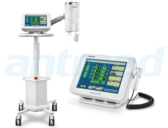
Our ANT000150 DSA Single Head Injector delivers contrast precisely in Digital Subtraction Angiography.
To start with, this injector ensures safety: A double confirmation mechanism for air purging and injection readiness avoids hazards and protects patients. It can automatically slow and stop at 1200 PSI pressure, with visual and aural alerts.
Besides, the color touchscreen is easy to operate; users can easily switch between the operating room and the scanning room. It also offers real-time pressure monitoring and optimal contrast temperature for predictable results. The rotating, waterproof injection head saves operational errors, while connectivity with other imaging systems improves procedural efficiency. Our design focuses on accuracy in crucial circumstances with a 0.1 to 45 ml/s flow rate and 2000 injection program memory.

Conclusion
Antmed specializes in high-pressure contrast imaging systems for CT, MRI, and DSA. Our high-pressure contrast injectors and syringes offer safety, accuracy, and interoperability with key imaging systems. We fulfill the changing medical demands of the globe with our patent portfolio and devotion to technological progress. If you are looking for high-quality contrast media injectors, visit Antmed’s website!
- Address : 18 Jinhui Ave., Pingshan District, Shenzhen, 518122, China
- Tel : +86-755-86060992
- Email : info@cdpaigao.com


 Medical Imaging Products
 Anesthesia Intensive Care
 Cardiovascular and Peripheral Products